Female Genital/Sexual Anatomy
Written by Dr.M.D.Mazumdar, MD
The Female Genital Organs consists of the external genital organs (or the 'vulva') and the internal genital organs. The female breasts are considered the accessory organs of reproduction.
Female External Genital Organs
The 'Vulva' or the female external genital organs, are those genital organs that are present on the surface of the female body and can be easily examined without the use of any special instruments. These are also the female sexual organs.
The Vulva consists of the mons pubis, the labia majora, the labia minor, clitoris, vestibule and glands like the Bartholin's glands, Skene's glands and the Vestibular Glands.

Female External Reproductive Anatomy
The perinuem - the area between the vulva and the anus - is also considered to be a part of the female external genital organs.
The other external genital organs are enclosed between the labia majora. They are the two thin labia minora, the clitoris, the vestibule, the external urethral meatus, the vaginal opening covered with a hymen in virgins and a number of glands like the Bartholin's glands and the vestibular glands.
Of these, the clitoris and the labia minora are the important sexual organs. (Read More ...)
Female Internal Genital Organs
The female internal genital organs can be examined only with the help of instruments.
The vagina is a hollow muscular canal which opens on on the external surface of the body between the two labia minora. Its other side is closed against the uterus. It can be examined with the fingers or with the help of simple instruments like the speculum. Although it is included among the internal organs, the lower part can also be seen partially from the outside - this is especially so in elderly women in whom the muscles become lax and the vaginal opening tends to gape open. In virgins, the vagina is partially closed by a thin membrane known as the hymen.

Female Internal Reproductive Anatomy
The vagina is the organ for sexual intercourse. The G-spot is an erogenous spot in the posterior wall of the vagina.
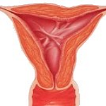
The other internal genital organs are the uterus, the two fallopian tubes and the two ovaries.
The uterus is a muscular, pear shaped organ which has a tremendous capacity to increase in size during pregnancy. The non-pregnant uterus is about 50-80 gms in weight and can hold only about 5 ml of fluid. But at full term pregnancy, it can weigh as much as 1 kg and hold not only the baby but also the placenta, the membranes, as well as a large amount of amniotic fluid.
The two Fallopian tubes extend form the uterus on either side to reach the ovaries. At the time of ovulation ( at around the 14th day after the period) the tubes pick up the ovum released from one of the ovaries. The ovum then moves through the tubes towards the uterus. Fertilization occurs inside the tubes and the embryo moves on to the uterus to get embedded here. (Read more about How Pregnancy Occurs here ...). The ovaries not only release a mature ovum at ovulation but also secretes the two main female hormones - estrogen and progesterone. ( Read more ... ).
The Female Breasts
The female breasts are also known as the accessory organs of reproduction.
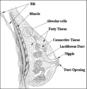
The Female Breast
While both men and women have breasts, it is the female breasts which increase in size during puberty under the influence of estrogen. Further size increase occurs during pregnancy. During pregnancy, the breasts also change in preparation for childbirth and breastfeeding.
The breasts essentially comprises of a mammary gland enclosed in layers of fat and fibrous tissue. The fibrous tissue helps in supporting the breasts.
There are about 15-20 lobes in each breast, each with a duct leading up to the nipple. Milk is produced in the glands, and is transported to the nipples by the ducts.
After the menopause, the glandular part of the breasts atrophy to some extent due to lack of stimulation by the low estrogen levels. The breast size is then maintained mainly by fat. ( Read More ...)
Read More:


Also Read-
- Caring for the Normal Pregnancy.
- Menstruation/Period and Why it Occurs.
- Vaginal Discharge with Itching .
- Causes and Treatment of Infertility.
Do you have a gynecological or obstetrical problem? Would you like to discuss it in private? Consult our online gynecologist Dr.M.D.Mazumdar, MD (O&G), at any time you want and get your reply within 24 hours.We charge a nominal fee of USD 20 ($20) per question through Paypal.com.
The procedure of asking a question is quite simple. Clicking on the link below takes you to the Paypal website where the payment is made. After the payment goes through, you will be directed back to this website where you can ask your question. And rest assured, you will get your answer within 24 hours. And usually, even sooner.